


Introduction: The Importance of Medical Imaging in Modern Healthcare
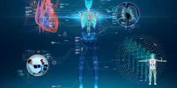
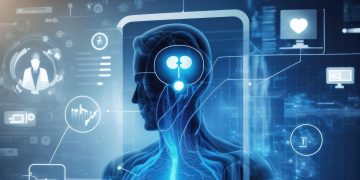
Medical imaging has revolutionized healthcare, enabling clinicians to peer inside the human body without resorting to invasive procedures. Over the past few decades, advances in medical imaging technologies have dramatically improved the ability to diagnose and treat a wide range of medical conditions. These technologies include X-rays, CT scans, MRI, ultrasound, and nuclear medicine. They provide crucial information about the structure and function of organs and tissues, allowing for more accurate diagnoses, better treatment planning, and improved patient outcomes.
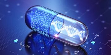
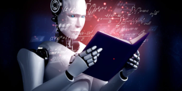
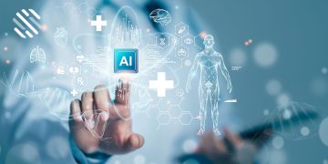
As the healthcare landscape evolves, artificial intelligence (AI) and machine learning (ML) are playing an increasingly prominent role in medical imaging. AI algorithms can now assist radiologists in interpreting images, automating tasks, and even identifying patterns that may not be immediately visible to the human eye. These advancements promise not only to enhance the accuracy of diagnoses but also to reduce the workload on medical professionals, enabling them to focus on patient care and complex cases.
In this article, we will explore the current state of medical imaging and its diagnostic applications, discuss emerging AI-powered diagnostic tools, and examine the challenges and future prospects of this technology.
1. The Evolution of Medical Imaging Technologies
1.1 Early Developments in Medical Imaging
The history of medical imaging began in 1895 when Wilhelm Roentgen discovered X-rays. This discovery enabled doctors to view the bones and internal structures of the body for the first time, transforming the diagnosis of fractures and other skeletal issues. The first X-ray machines were rudimentary, but over time, the technology evolved, leading to the creation of more sophisticated imaging modalities.
1.2 Advancements in Imaging Modalities
Since the introduction of X-rays, several other imaging techniques have been developed, each with its own advantages and specific applications in diagnosis:
- Computed Tomography (CT): CT scans combine X-rays with computer processing to create detailed cross-sectional images of the body. This imaging technique is particularly useful for identifying tumors, internal injuries, and vascular abnormalities.
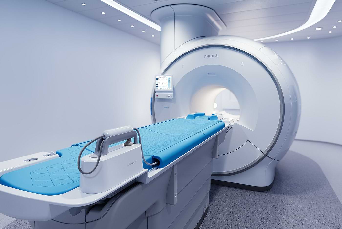
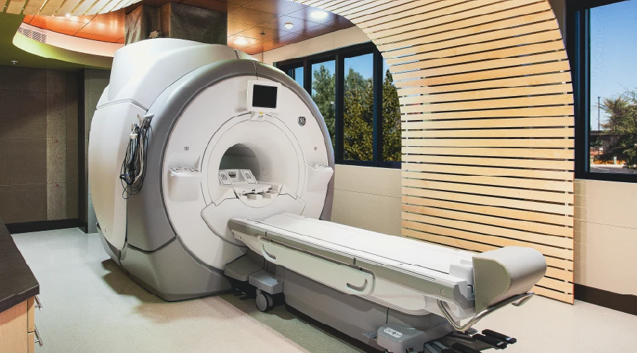
- Magnetic Resonance Imaging (MRI): MRI uses strong magnetic fields and radio waves to generate detailed images of soft tissues, making it ideal for imaging the brain, spinal cord, muscles, and joints. Unlike X-rays, MRI does not use ionizing radiation, making it a safer option for certain patient populations.
- Ultrasound: Ultrasound imaging uses high-frequency sound waves to produce images of the internal organs, muscles, and tendons. It is commonly used in obstetrics (e.g., monitoring pregnancies), cardiology (e.g., assessing heart function), and emergency medicine (e.g., evaluating trauma injuries).
- Positron Emission Tomography (PET): PET scans use radioactive tracers to create images that show how organs and tissues are functioning. This modality is frequently used in oncology to detect cancer and monitor treatment responses.
- Single Photon Emission Computed Tomography (SPECT): SPECT, similar to PET, uses radioactive tracers to evaluate the function of organs and tissues. It is commonly used for cardiac imaging, neurological evaluations, and cancer diagnostics.
These imaging techniques have dramatically increased the speed and accuracy of medical diagnoses, contributing to early detection and timely intervention for a wide range of diseases and conditions.
2. The Role of AI in Medical Imaging
2.1 AI’s Growing Influence in Healthcare
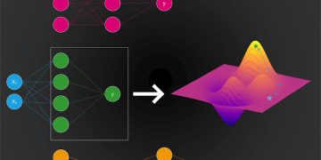
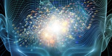
Artificial intelligence is transforming the healthcare industry, and medical imaging is no exception. Machine learning (ML) algorithms, particularly deep learning (DL), are now being used to assist in image analysis, offering significant advantages over traditional manual methods. By training algorithms on large datasets of medical images, AI models can learn to identify patterns, detect abnormalities, and make predictions that help radiologists arrive at more accurate diagnoses.
2.2 AI-Assisted Image Interpretation
One of the most significant applications of AI in medical imaging is automated image interpretation. AI-powered tools can help radiologists interpret images more accurately and efficiently by detecting potential issues in medical scans that might otherwise go unnoticed. Some specific examples include:
- Tumor Detection: AI algorithms are trained to identify potential tumors, lesions, or nodules in CT, MRI, and X-ray images, significantly improving early detection rates for cancers such as breast, lung, and brain cancer.
- Fracture Identification: AI models can analyze X-ray images to quickly detect fractures, even in complex or challenging areas of the body. This feature is particularly useful in emergency departments where radiologists may be overwhelmed by a large volume of images.
- Cardiovascular Disease Detection: AI tools are being employed to analyze echocardiograms, CT angiograms, and other cardiac images to identify conditions like coronary artery disease or heart valve abnormalities.
- Pulmonary Disease Diagnosis: AI models can assess chest X-rays and CT scans for signs of pneumonia, pulmonary embolism, chronic obstructive pulmonary disease (COPD), and even the effects of COVID-19 on the lungs.
2.3 AI in Image Reconstruction and Enhancement
In addition to aiding diagnosis, AI is also improving the quality of medical images through reconstruction and enhancement techniques. For example, AI algorithms can be used to reduce noise and improve image resolution, resulting in clearer and more detailed images. This enhancement improves the accuracy of diagnoses and minimizes the need for additional imaging procedures.
AI can also play a key role in image segmentation, where the algorithm isolates specific structures or regions of interest, such as tumors, blood vessels, or organs, from the rest of the image. This is particularly important in treatment planning, as it allows for precise targeting during procedures like radiation therapy.
2.4 Real-Time Diagnostics
AI-driven systems are also making it possible to perform real-time diagnostics. For instance, AI can process images immediately after they are taken, providing clinicians with quick feedback on potential issues. This is particularly useful in emergency settings, where time is critical, and in telemedicine, where remote diagnosis is increasingly common.
3. AI in Diagnostic Assistance: Improving Efficiency and Accuracy
3.1 Automating Routine Tasks
AI’s ability to automate routine tasks in medical imaging not only saves time but also reduces the cognitive load on radiologists and other healthcare professionals. Tasks such as image preprocessing, triaging, and prioritization can be automated, allowing clinicians to focus on more complex cases that require human expertise. AI systems can flag high-priority cases, such as potential cancer diagnoses, and alert doctors for immediate attention.
3.2 Predictive Analytics and Early Detection
One of the most exciting aspects of AI-assisted medical imaging is the ability to perform predictive analytics. By analyzing large volumes of medical images along with patient data (e.g., genetics, medical history, lifestyle factors), AI can help predict disease outcomes and treatment responses. This can lead to earlier interventions, better prognostic predictions, and the ability to tailor treatments to individual patients.
For example, in the case of breast cancer, AI models can predict the likelihood of metastasis based on the appearance of mammograms and biopsy results. Similarly, AI can help predict the progression of neurodegenerative diseases like Alzheimer’s disease by analyzing MRI scans of the brain.
3.3 Enhancing Collaboration and Decision-Making
AI also facilitates collaboration among healthcare providers. With the integration of AI tools, clinicians can quickly share and discuss imaging results across different departments, improving communication and decision-making. For instance, radiologists, oncologists, and surgeons can collaborate more efficiently in developing treatment plans based on AI-assisted imaging results.
4. Challenges and Limitations
4.1 Data Quality and Diversity
The effectiveness of AI in medical imaging relies heavily on the quality and diversity of the training data. AI models must be trained on large, high-quality datasets that represent a wide range of patient demographics, medical conditions, and imaging techniques. Without diverse data, AI models risk overfitting or producing biased results that are not generalizable to all patient populations.
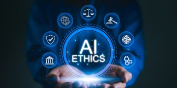
4.2 Regulatory and Ethical Concerns
The adoption of AI in medical imaging is subject to regulatory oversight to ensure patient safety. Regulatory bodies such as the FDA (U.S. Food and Drug Administration) must evaluate and approve AI systems before they can be used in clinical practice. Furthermore, the ethical implications of AI in healthcare, including data privacy, informed consent, and algorithmic fairness, must be carefully considered.
4.3 Integration into Clinical Workflow
Although AI offers significant potential, integrating these technologies into the existing clinical workflow can be challenging. Resistance to change, training requirements, and the need for robust cybersecurity measures are all factors that need to be addressed to ensure the smooth integration of AI tools into hospitals and clinics.
5. The Future of Medical Imaging and Diagnostic Assistance
As AI continues to evolve, the future of medical imaging and diagnostic assistance looks promising. The integration of AI into medical imaging systems is expected to enhance diagnostic accuracy, reduce clinician workload, and improve patient outcomes. With the continued development of AI algorithms and imaging technologies, we can expect even greater precision in diagnosis, faster response times, and a more personalized approach to patient care.
Key developments on the horizon include:
- Improved AI Algorithms: The next generation of AI models will be able to analyze more complex and varied data, further improving diagnostic capabilities.
- Personalized Imaging: AI will allow for more personalized imaging protocols tailored to an individual’s specific health profile, improving the effectiveness of diagnostic imaging.
- Integration with Other Technologies: AI-driven diagnostic tools will be increasingly integrated with other medical technologies, such as robotic surgery, electronic health records (EHR), and telemedicine, offering a comprehensive solution for patient care.
Conclusion
Medical imaging has been a cornerstone of modern healthcare, and the integration of artificial intelligence has taken its capabilities to unprecedented levels. By automating interpretation, enhancing image quality, and providing predictive analytics, AI is poised to revolutionize diagnostic practices across the globe. While there are challenges to overcome, the future of AI in medical imaging is bright, with the promise of more accurate, efficient, and personalized healthcare for patients worldwide.
As these technologies continue to advance, medical professionals and AI developers must work together to ensure that the benefits of AI are realized in a way that prioritizes patient safety, privacy, and care quality. The collaboration between human expertise and AI is set to define the next era of diagnostic medicine, leading to better outcomes and enhanced healthcare delivery.


